Fayl:DTI-sagittal-fibers.jpg
Sınaq göstərişi ölçüsü: 643 × 600 piksel. Digər ölçülər: 257 × 240 piksel | 515 × 480 piksel | 1.021 × 952 piksel.
Faylın orijinalı (1.021 × 952 piksel, fayl həcmi: 294 KB, MIME növü: image/jpeg)
|
Xülasə
| İzah |
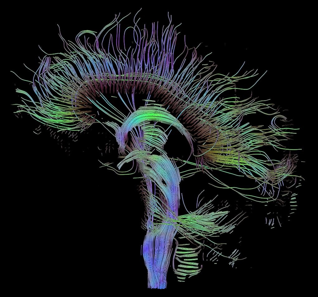
English: Visualization of a DTI measurement of a human brain. Depicted are reconstructed fiber tracts that run through the mid-sagittal plane. Especially prominent are the U-shaped fibers that connect the two hemispheres through the corpus callosum (the fibers come out of the image plane and consequently bend towards the top) and the fiber tracts that descend toward the spine (blue, within the image plane)
Français : Visualisation d'une mesure DTI d'un cerveau humain. Ce qui est représenté sont des faisceaux de fibres reconstruits qui traversent le plan demi-sagittal. On observe les fibres en U qui connectent les deux hémisphères à travers le corps calleux, qui sont particulièrement importantes (les fibres sortent du plan de l'image et par conséquent se courber vers le haut) ainsi que les faisceaux de fibres qui descendent vers la colonne vertébrale (bleu, dans le plan de l'image)
Deutsch: Traktographie-Verfahren rekonstruieren aus den Messdaten der Diffusions-Tensor-Bildgebung den anzunehmenden Verlauf größerer Nervenbahnen. Hier dargestellt sind die Ergebnisse für ein menschliches Gehirn; um die Übersichtlichkeit zu wahren, beschränkt sich die Abbildung auf Bahnen, die die Medianebene schneiden. Insbesondere sind dies die U-förmigen Faserbündel, die die beiden Hirnhälften verbinden (sie durchstoßen die Bildebene und sind nach oben gebogen) sowie die Faserbündel, die zum Rückenmark ziehen (blau dargestellt, liegen innerhalb der Bildebene) |
| Tarix | |
| Mənbə | Öz işi |
| Müəllif | Thomas Schultz |
| İcazə (Faylın təkrar istifadəsi) |
Rendering is own work, using a modified version of the BioTensor application developed at the University of Utah. The dataset is courtesy of Gordon Kindlmann at the Scientific Computing and Imaging Institute, University of Utah, and Andrew Alexander, W.M. Keck Laboratory for Functional Brain Imaging and Behaviour, University of Wisconsin, Madison. It is publicly available from [1] |
Lisenziya
I, the copyright holder of this work, hereby publish it under the following licenses:

|
Bu sənədi GNU Azad Sənədləşdirmə Lisenziyası, Versiya 1.2 və ya Azad Proqram Fondu tərəfindən nəşr olunan hər hansı sonrakı versiya şərtlərinə əsasən dəyişməz bölmələr, ön qapaq mətnləri və arxa qapaq mətnləri olmadan köçürmək, yayımlamaq və / və ya dəyişdirmək üçün icazə verilir; Lisenziyanın bir nüsxəsi GNU Azad Sənədləşdirmə Lisenziyası adlı hissəyə daxil edilmişdir. |
| Bu fayl Creative Commons Attribution-Share Alike 3.0 Unported lisenziyası altında yayımlanır. | ||
| ||
| This licensing tag was added to this file as part of the GFDL licensing update. |
This file is licensed under the Creative Commons Attribution-Share Alike 2.5 Generic, 2.0 Generic and 1.0 Generic license.
- Azadsınız:
- paylaşmaq – əsəri köçürmək, paylamaq və ötürmək üçün
- remiks etmək – əsəri adaptasiya etmək
- Aşağıdakı şərtlərə riayət etməklə:
- istinad – Müvafiq kredit verməlisiniz, lisenziyaya bir keçid verməlisiniz və dəyişikliklərin olub olmadığını bildirməlisiniz. Bunu hər hansı bir ağlabatan şəkildə edə bilərsiniz, ancaq lisenziyalaşdırıcının sizi və ya istifadənizi təsdiqləməsini təklif edən bir şəkildə deyil.
- bənzər paylaşma – Əsəri remix edirsinizsə, dəyişdirirsinizsə və ya üzərində iş aparırsınızsa, öz töhfələrinizi orijinalda olduğu kimi eyni və ya uyğun lisenziya altında yayımlamalısınız.
İstədiyiniz lisenziyanı seçə bilərsiniz.
Faylın tarixçəsi
Faylın əvvəlki versiyasını görmək üçün gün/tarix bölməsindəki tarixlərə klikləyin.
| Tarix/Vaxt | Kiçik şəkil | Ölçülər | İstifadəçi | Şərh | |
|---|---|---|---|---|---|
| indiki | 10:42, 13 oktyabr 2017 | | 1.021 × 952 (294 KB) | Mikael Häggström | Minor crop of black areas at the top and bottom |
| 16:22, 22 sentyabr 2006 |  | 1.021 × 1.125 (203 KB) | Thomas Schultz | {{Information |Description=Visualization of a DTI measurement of a human brain. Depicted are reconstructed fiber tracts that run through the mid-sagittal plane. Especially prominent are the U-shaped fibers that connect the two hemispheres through the corp |
Fayl keçidləri
Aşağıdakı səhifə bu faylı istifadə edir:
Faylın qlobal istifadəsi
Bu fayl aşağıdakı vikilərdə istifadə olunur:
- af.wikipedia.org layihəsində istifadəsi
- ar.wikipedia.org layihəsində istifadəsi
- bn.wikipedia.org layihəsində istifadəsi
- cs.wikipedia.org layihəsində istifadəsi
- de.wikipedia.org layihəsində istifadəsi
- Autismus
- Computergrafik
- Bipolare Störung
- Portal:Informatik/Exzellente Artikel
- Portal:Geist und Gehirn/Artikel des Monats
- Diffusions-Tensor-Bildgebung
- Wikipedia:Kandidaten für exzellente Bilder/Archiv2006/17
- Datei:DTI-sagittal-fibers.jpg
- Wikipedia:Exzellente Bilder/Naturwissenschaften
- Portal:Physik/Artikel des Monats 2024-03
- Wikipedia:Exzellente Bilder/Kleine Bilder
- en.wikipedia.org layihəsində istifadəsi
- Neurolinguistics
- Tractography
- Portal:Medicine
- User talk:Spikebrennan
- User:Spikebrennan
- Diffusion MRI
- Wikipedia:WikiProject Neuroscience
- Portal:Psychology/Selected article
- Wikipedia:Featured pictures/Sciences/Biology
- Portal:Psychology/Selected article/7
- Wikipedia:Featured pictures thumbs/08
- Wikipedia:Featured picture candidates/DTI-sagittal-fibers.jpg
- Wikipedia:Wikipedia Signpost/2007-11-05/Features and admins
- Wikipedia:Featured picture candidates/November-2007
- Wikipedia:Picture of the day/March 2008
- Connectome
- Template:POTD/2008-03-10
- User talk:Thomas Schultz
- Wikipedia:Wikipedia Signpost/2007-11-05/SPV
- Biological data visualization
- Wikipedia:WikiProject Medicine/Recognized content
- Wikipedia:WikiProject Molecular Biology/Biophysics
- User:Wouterstomp/test
- Wikipedia:WikiProject Anatomy/Resources
- Wikipedia:WikiProject Anatomy/Recognized content
- Wikipedia talk:WikiProject Anatomy/Archive 9
- Portal:Medicine/Recognized content
- User talk:Rhododendrites/Reconsidering FPC on the English Wikipedia
- User:Hydrogenkitsch
- Wikipedia:Wikipedia Signpost/Single/2007-11-05
- en.wikibooks.org layihəsində istifadəsi
- en.wikiquote.org layihəsində istifadəsi
Bu faylın qlobal istifadəsinə baxın.